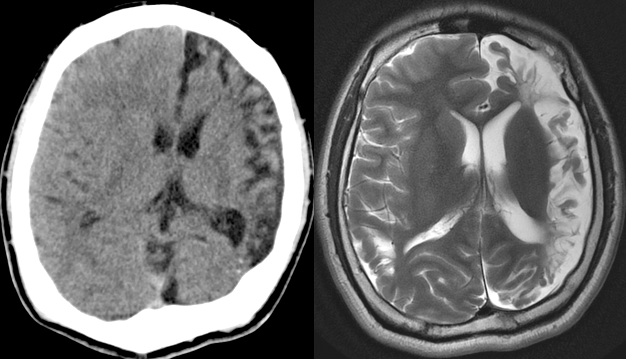
[Left]: Head CT shows left hemispheric volume loss. The injury happened early enough that even the skull is smaller on that side.
[Right]: Brain MRI shows the severe left hemispheric atrophy. Some of the brain gyri have bulbous ends and a thin neck, resembling mushrooms, a shape called ulegyria and consequence of the brain atrophy. The left lateral ventricle is mildly enlarged due to the atrophied brain.
3 Comments
gg123456789@lemmy.world · 1 pts · 3y
Any ideas which sequence the MRI image comes from (I'm guessing MP2RAGE but I'm seeing more vessels like FLAIR)?
Spectator@lemmy.world · 1 pts · 3y
Just a standard T2 sequence.
FLAIR would have dark CSF, since that's what FLAIR is designed to suppress - CSF.
MPRAGE is a T1 sequence that's usually done with contrast.
gg123456789@lemmy.world · 1 pts · 3y
Got it, thanks for clarifying 👍